|
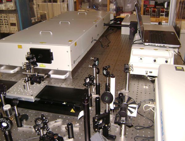
DescriptionSpectra-Physics: Amplifier Spitfire Ace (5mJ, 35fs, 800nm, 1kHz) ; Optical Parametric Amplifier (Topas, 290nm-2600 nm); Research:
Materials:
Contact person: dr hab. Andrzej Stupakiewicz prof. UwB, E-mail:and@uwb.edu.pl Scanning Probe Microscope Solver HV+ Ntegra Prima NT-MDT
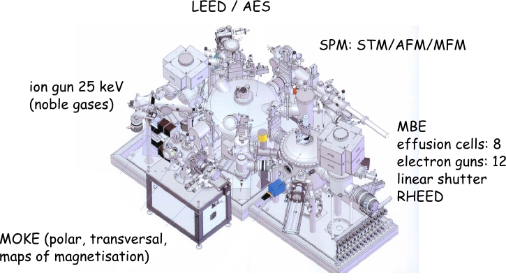
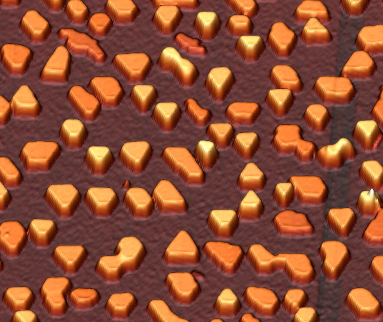
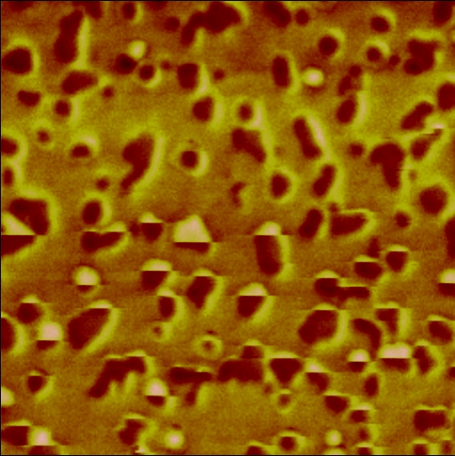
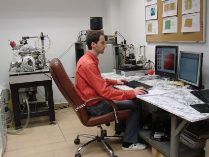
Scanning Probe Microscope is equipped with electro-magnets system (either in-plane magnetic field up to 0.2T or perpendicular field up to 0.1T), cold finger system (110K-450K), vacuum chamber (10-6 Torr), vibration isolation system and videomicroscope with manual continuous zoom. Study of surface topography as well as magnetic, electrical and electrochemical properties is possible. Force and voltage lithography is available. Measurements can be done in both high vacuum and in air (contact AFM, LFM, Resonant Mode (semicontact + noncontact AFM), Phase Imaging, Force Modulation (viscoelastisity), MFM, EFM, Adhesion Force Imaging, AFM Litography-Force, Spreading Resistance Imaging ("conductive" AFM), AFM Litography-Voltage, Scanning Capacitance Imaging, Scanning Kelvin Microscopy) as well as in a liquid (contact AFM, LFM, Adhesion Force, Force Modulation (viscoelastisity), Resonant Mode (semicontact AFM), Phase Imaging, AFM Litography-Force). Contact person: mgr Piotr Mazalski, e-mail:piotrmaz@uwb.edu.pl High Sensitivity Magneto-Optical Kerr Magnetometer
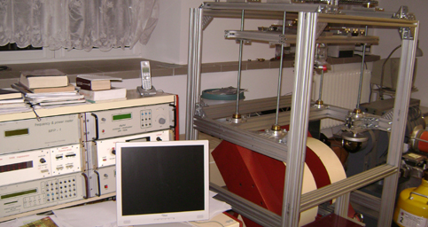
High Sensitivity Magneto-Optical Kerr Magnetometer equipped with special electro-magnets system (perpendicular to the plane sample up to 20kOe and in-plane up to 4kOe magnetic field), Coherent Ar (Innova 300), dye (599-2) and red-diode lasers, optical cryostat (10-350K), photo-elastic modulator (s,p-polarization, rotation and ellipticity polar, longitudinal, transverse Kerr effects, linear and circular dichroism). Study of static and dynamics magnetization reversal processes and vector magnetization analysis in ultrathin multilayer magnetic films with specialized LabView software for control magnetometer and data analysis. Contact person: dr hab. Andrzej Stupakiewicz prof. UwB, E-mail:and@uwb.edu.pl Magnetometers based on optical polarizing microscopes
|
|
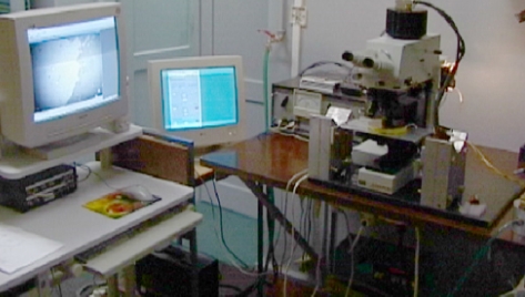
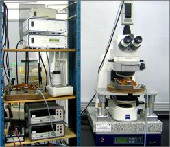
Laboratory is equipped with self-constructed optical systems and SPINLAB supported bought microscope system.
- Carl Zeiss Jenapol optical polarizing Kerr microscope
- special electro-magnets (out-of-plane or in-plane, static and pulse magnetic field),
- Carl Zeiss Xe and halogen lamps;
- high sensitivity 12-bit MicroMax CCD camera.
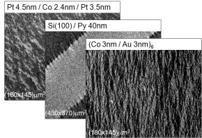
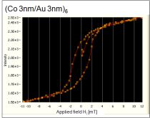
Study of statics and dynamics of domain structure (mainly in nanostructures) is possible by the structure imaging with both perpendicular and in-plane magnetization components. Specialized image processing techniques and LabView software for control and data analysis were developed.
![]() SPINLAB supported system
SPINLAB supported system
 |
 |
|
|
 |
Contact person: dr hab. Maria Tekielak, E-mail:tekmar@uwb.edu.pl
Keywords: magneto-optical Kerr effect, magnetic domain structure, static and dynamics magnetic domain wall
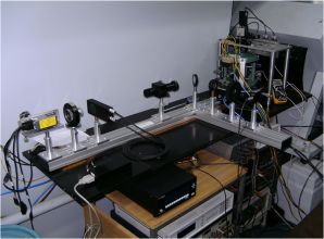
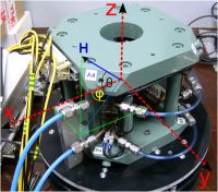
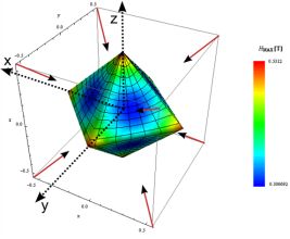
3D magnetic field system for magnetooptic investigations 
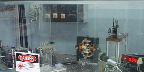
3D magnetic field system for magnetooptic investigations.
![]() SPINLAB supported system
SPINLAB supported system
 |
 |
|
|
 |
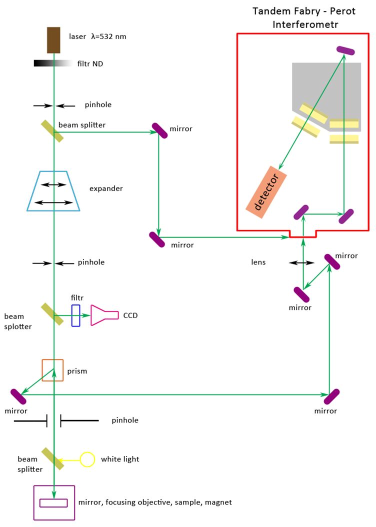
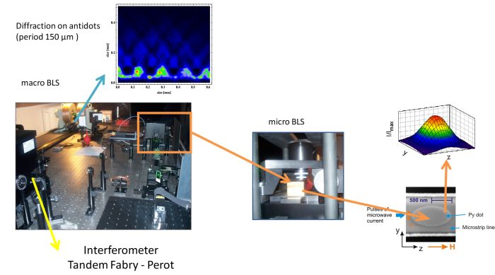
Time and space resolved Brillouin light scattering system 
![]() SPINLAB supported system
SPINLAB supported system
Cryogen free systems for magnetooptical investigations 
![]() SPINLAB supported system
SPINLAB supported system
|
Closed-cycle, cryo-free cryostat DE-204 PF Advanced Research System, Inc, USA, for magnetooptical-Kerr-microscopy-investigation of spatial distribution of magnetization.
|
Closed-cycle, cryo-free cryostat CFSM7T-1.5 OXFORD INSTRUMENTS PLC for magnetooptical investigation of ultra-fast magnetization processes in low temperatures and high magnetic field.
|
Ferromagnetic resonance spectrometer
|
Short description
A high - sensitivity ferromagnetic resonance spectrometer at a microwave frequency of 9.5 GHz and at room temperatures with computerized data collection systems for magnetic anisotropy analysis.
Full description
A high - sensitivity ferromagnetic resonance spectrometer at a microwave frequency of 9.5 GHz and room temperatures with computerized data collection systems. The FMR spectra are investigated as a function of the orientation of the applied dc field. Study of magnetic anisotropy in thin and super thin magnetic samples.
Contact person: dr. Ryszard Gieniusz, E-mail:gieniusz@uwb.edu.pl
Keywords:Ferromagnetic resonance, magnetic anisotropy, magnetic films.
 |
 |
 |
